Finding related functional neuroimaging volumes
Finn Årup Nielsen and Lars Kai Hansen
Running title: Finding related volumes
Keywords: Functional neuroimaging, information retrieval,
content-based image retrieval, multivariate analysis.
Address for correspondence:
Finn Årup Nielsen
Informatics and Mathematical Modelling, Building 321,
Technical University of Denmark, Lyngby, Denmark
Tel: +45 4525 3921
Fax: +45 4587 2599
E-mail: fn@imm.dtu.dk
Abstract:
We describe a content-based image retrieval technique for finding
related functional neuroimaging experiments by voxelization of sets of
stereotactic coordinates in Talairach space, comparing the volumes and
reporting related volumes in a sorted list.
Voxelization is accomplished by convolving each coordinate with a
Gaussian kernel.
The scheme allows us to compare experiments represented as either
lists of coordinates or volumes, and we introduce alternative
entrances to databases by image-based indices constructed via novelty
measures and singular value decomposition.
Identification of related research in functional neuroimaging can be
carried out, e.g., by searching in bibliographic databases such as PubMed,
browsing ``table of contents'' of scientific journals or searching
BrainMap [Fox and Lancaster, 1994] with, e.g., behavioral or location
criteria.
Here we describe a content-based image retrieval method based on
activation information in 3-dimensional (3D) Talairach space
[Talairach and Tournoux, 1988].
The information might either come in the form of a list of points
representing activation hot spots or it might come as a statistical
parametric map, e.g., a volume of  -statistics from a statistical
analysis of a functional neuroimaging data set.
Our first goal is to establish a service comparable to ``Related
Articles'' of PubMed.
-statistics from a statistical
analysis of a functional neuroimaging data set.
Our first goal is to establish a service comparable to ``Related
Articles'' of PubMed.
Retrieval systems for digital text have existed for several decades
and are often based on the vector space model [Salton, 1971],
where a document is represented in a vector with each element
associated with a word or phrase.
Retrieval systems for other digital objects than text
have also been proposed, e.g, on images and sounds
[Feiten and Günzel, 1994].
Some image retrieval systems are based on text description of images,
but others have included features, e.g.,
color, texture, shape and keywords.
This allows for image query by example, i.e., ``Show me images similar
to this image'' as in the IBM QBIC (Query by Image Content) system
[Flickner et al., 1995,Faloutsos et al., 1994,Niblack et al., 1993].
A number of other systems exists, see [Eakin, 2000] for
a list.
Web-based image retrieval systems have also been suggested
[Sclaroff, 1995] and implemented in, e.g., WebSEEk
[Smith and Chang, 1996] and ImageRover
[Sclaroff et al., 1997] as well as AltaVista
(http://www.altavista.com).
Neuroimaging retrieval systems have also been constructed, e.g.,
[Liu and Dellaert, 1998]
describe image retrieval for 3D medical images specifically CT brain
scans containing normal, stroke and ``blood cases''.
The basic ``object'' is a half 2D slice where features are extracted
from, such as mean, standard deviation and asymmetry measures.
Other medical image retrieval systems and methods have been described,
e.g.,
[Petrakis and Faloutsos, 1997], [Chu et al., 1998],
I net [Orphanoudakis et al., 1996],
MIMS [Chbeir et al., 1999] and
a system for decision support in clinical pathology
[Comaniciu et al., 1999].
net [Orphanoudakis et al., 1996],
MIMS [Chbeir et al., 1999] and
a system for decision support in clinical pathology
[Comaniciu et al., 1999].
In research-oriented functional neuroimaging retrieval systems the
BrainMap stands out:
BrainMap is a database holding functional neuroimaging
studies [Fox and Lancaster, 1994] both accessible through a
web-interface and a stand-alone program
[Lancaster et al., 1997].
It allows for search via ``reference'', ``behavioral'', ``location''
and ``protocol'' criteria.
A location criterion can consist of a bounding box in Talairach space.
Finding related volumes was also considered in
[Van Horn et al., 2001] in connection with the Functional Magnetic
Resonance Data Center,
though this database at the time of writing implements search via
the bibliographic information only,
and [Ford et al., 2001] describe briefly an ``inter- and
intra-study data mining'' tool for functional magnetic resonance
imaging (fMRI) activation maps
based on ``activation signatures'' such as size, shape, number of
foci, location and orientation.
A related method identifies global patterns and ``cluster''
experiments [Lloyd, 2000]: Multidimensional scaling was used
to map 35 positron emission tomography (PET) studies to a
3D space based on their activations represented in an
87-dimensional space redundantly
comprising Brodmann areas, gyri, sulci and lobes.
Earlier, brief descriptions of our work are available in
[Nielsen, 2001,Nielsen and Hansen, 2002a].
We downloaded the BrainMap database through its web-site and extracted
fields that were relevant for our purpose.
Since its activation data is in an ``experiment'' structure containing
a variable length list of activation foci (``locations'') we
convert the 3D locations to a voxel-volume representation by a voxelization step where each location  in an
``experiment'' is convolved with a Gaussian
kernel in the same manner as a Parzen window/Specht
kernel estimation
[Nielsen and Hansen, 2002b,Turkeltaub et al., 2002].
Some of the locations carry a sign and in the present application we
maintain this sign and negate the kernel for the negative locations.
For those voxels where the sign is not explicit we assume that it is
positive.
We normalize with the number of locations in each experiment, thus if
there are no negative locations the voxelized volume is a probability
density volume.
The full voxelization equation determining the value
in an
``experiment'' is convolved with a Gaussian
kernel in the same manner as a Parzen window/Specht
kernel estimation
[Nielsen and Hansen, 2002b,Turkeltaub et al., 2002].
Some of the locations carry a sign and in the present application we
maintain this sign and negate the kernel for the negative locations.
For those voxels where the sign is not explicit we assume that it is
positive.
We normalize with the number of locations in each experiment, thus if
there are no negative locations the voxelized volume is a probability
density volume.
The full voxelization equation determining the value  at the voxel
 is from
  locations
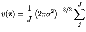 sgn sgn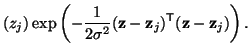 |
(1) |
We fix the kernel width at  mm corresponding to
approximately 24 mm full width half maximum.
This width should incorporate both the uncertainty of the location as
well as the spatial extent of the original activation
[Brett et al., 2002].
Due to memory constraints we use a coarse sampling with
mm corresponding to
approximately 24 mm full width half maximum.
This width should incorporate both the uncertainty of the location as
well as the spatial extent of the original activation
[Brett et al., 2002].
Due to memory constraints we use a coarse sampling with
 mm
mm voxel-sizes.
Voxelization can be regarded as the inverse operation of finding a
local maxima or the identification of the
center of gravity/mass of a connected region in a thresholded
volume.
voxel-sizes.
Voxelization can be regarded as the inverse operation of finding a
local maxima or the identification of the
center of gravity/mass of a connected region in a thresholded
volume.
Once all  volumes are constructed we vectorize each volume into a
 -length vector
![$ {\bf x}_n = [ v({\bf z}^n_1), \ldots, v({\bf z}^n_P)]$](img14.gif) and collect all vectors in a matrix
and collect all vectors in a matrix
![$ {\bf X}(N\times P) = [ {\bf x}_1, \ldots, {\bf x}_N ]^{\sf T}$](img15.gif) .
A similarity matrix
.
A similarity matrix  is computed as a normalized inner product
between the vectors
is computed as a normalized inner product
between the vectors
This measure is related to the reproducibility index in the NPAIRS framework
[Strother et al., 2002].
The similarities are sorted and for each volume the most similar
and most dissimilar volumes are reported in two lists.
Static HTML web-pages are generated containing both lists as well as
summaries of the experiment, a simple Corner Cube visualization
[Rehm et al., 1998] and links to BrainMap and Pubmed.
We further included six volumes from a motor learning positron
emission tomography (PET) study
[Balslev et al., 2002].
These volumes represent the cluster centers of a K-means
clustering [MacQueen, 1967,Goutte et al., 1999].
They were resampled and converted from MNI to Talairach space with
Brett's transformation [Brett, 1999].
The complete pipeline for both the volume data and the BrainMap data
is displayed in figure 1.
Figure 1:
Pipeline for finding related volumes for data from the
BrainMap database.
 |
Apart from indices based on the bibliographic information associated
with an experiment we can produce image-based indices.
A simple ad hoc novelty/outlier measure  is generated by
finding the mean volume
is generated by
finding the mean volume
 as
the average across all volumes and comparing this through the inner
product with all the volumes.
The novelty for the
as
the average across all volumes and comparing this through the inner
product with all the volumes.
The novelty for the  'th experiment is returned as the absolute value
of the inverse normalized inner product
'th experiment is returned as the absolute value
of the inverse normalized inner product
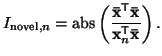 |
(3) |
See [Nielsen and Hansen, 2002b] for more advanced outlier detection in
neuroinformatics.
An other image-based index is generated through singular value
decomposition (SVD) of the experiment  voxel matrix
  , --
related to principal component analysis (PCA) as used for PET
and fMRI [Friston et al., 1993,Hansen et al., 1999]
, --
related to principal component analysis (PCA) as used for PET
and fMRI [Friston et al., 1993,Hansen et al., 1999]
 svd svd |
(4) |
For this operation we only include entries from the BrainMap database
that have ``Peer Reviewed'' as publication type, excluding reviews and
unpublished studies that would otherwise contribute with a
considerable part of the variance in .
We compare the 20 first eigenimages in  with each individual
volume in
with each individual
volume in  by a simple inner product and construct two lists
for each eigenimage:
One with the volumes that are most similar with the eigenimage and a
second with volumes that are most dissimilar (or similar to the
eigenimage with all signs reversed).
Both lists are equally important since the sign on an eigenimage
by a simple inner product and construct two lists
for each eigenimage:
One with the volumes that are most similar with the eigenimage and a
second with volumes that are most dissimilar (or similar to the
eigenimage with all signs reversed).
Both lists are equally important since the sign on an eigenimage
 can be reversed if the sign of
  is also changed.
We expect that the eigenimages will correspond to global
patterns within the entire set of studies.
is also changed.
We expect that the eigenimages will correspond to global
patterns within the entire set of studies.
As a small test we compared two extra studies
[Hyder et al., 1997,Phelps et al., 1997] to the data set from the
BrainMap database.
These two studies are fMRI reproductions of PET studies investigating
a ``willed action'' component with a sensorimotor and a verbal task
[Frith et al., 1991].
We should expect the corresponding volumes to appear high in the list
of related volumes.
[Hyder et al., 1997,Phelps et al., 1997] have restricted field of view
only covering the frontal part of the brain.
The tools for this analysis are implemented in the Brede
toolbox [Nielsen and Hansen, 2000] available from
http://hendrix.imm.dtu.dk/software/brede/ and the resulting web-pages
with volumes are presently available from
http://hendrix.imm.dtu.dk/services/jerne/.
Figure 2:
Example view of the related volumes for an experiment
reported in [Corbetta et al., 1993] and available in the
BrainMap database.
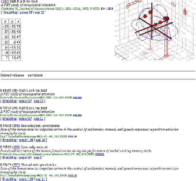 |
797 HTML pages were generated and the voxelized volumes consisted of
7752 voxels.
An example of one of the generated web-pages is displayed in figure 2
based on one of the 12 experiments/volumes reported in
[Corbetta et al., 1993].
It shows two clusters of activations: one in the parietal lobe and an
other in the frontal lobe.
This pattern is repeated for the five most related experiments.
Figure 3:
Novelty index showing a list of the most novel
experiments.
 |
The top of the novelty index is shown in figure 3:
The highest novelty is recorded in one of the three experiments
of [Allison et al., 1994].
The paper is the only one recorded with the ``electrophysiological''
modality (through implanted electrodes and combined with MRI).
Only  and
and  Talairach coordinates are shown in the
article, and the
Talairach coordinates are shown in the
article, and the  -coordinate in the database have been
estimated during entry.
Its high novelty might be due to this estimation and the rare modality.
The second largest novelty for a ``Peer Reviewed'' experiment appears
for the 5th entry in the table: [Reiman et al., 1989]
finds activation in the temporal pole in connection with anticipatory
anxiety.
A correction to this article later appeared where it was found that
the activation might not be a brain activation but an extracranial
muscle ``activation'' from teeth-clenching [Drevets et al., 1992].
The third highest novelty for a peer reviewed experiment is our
cluster volume described in [Balslev et al., 2002] and it is
confined to the rim of the brain, and which we attributed to
a possible motion artifact.
-coordinate in the database have been
estimated during entry.
Its high novelty might be due to this estimation and the rare modality.
The second largest novelty for a ``Peer Reviewed'' experiment appears
for the 5th entry in the table: [Reiman et al., 1989]
finds activation in the temporal pole in connection with anticipatory
anxiety.
A correction to this article later appeared where it was found that
the activation might not be a brain activation but an extracranial
muscle ``activation'' from teeth-clenching [Drevets et al., 1992].
The third highest novelty for a peer reviewed experiment is our
cluster volume described in [Balslev et al., 2002] and it is
confined to the rim of the brain, and which we attributed to
a possible motion artifact.
Figure 4:
The isosurfaces from the
positive and the negative end of the second eigenimage.
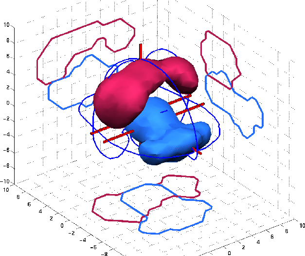 |
Since the mean of the images is not extracted our first eigenimage
from the SVD separates experiments with positive and
negative activations.
The top of the positive end contains among others two experiments by
[Parsons et al., 1995] each which contain 59 locations distributed
across large parts of the brain.
At the other end is an experiment [Silbersweig et al., 1993]
with 12 negative activations.
The subsequent eigenimages show high loadings on specific regions, e.g.,
the positive part of the second eigenimage covers the central sulcus
and nearby areas with a large weight on the left hemisphere, see
figure 4.
This implies that motion studies score high.
The other end of the eigenimage shows a loading in the occipital lobe
with the top experiments all involving visual stimulation, e.g., a
passive movement observation versus imaging grasping objects contrast
from [Decety et al., 1994] scores highest.
The next principal component distinguishes between cognitive and
sensorimotor experiments.
Higher components relates, e.g., to auditory presentation of words or
(visuo-)spatial processing.
Yet higher eigenimages show increasing spatial frequency and are harder
to interpret.
When the sensorimotor experiments of [Hyder et al., 1997] and
[Frith et al., 1991] are compared then [Hyder et al., 1997]
is found as the 4th most related of published studies in the list of
[Frith et al., 1991], and [Frith et al., 1991] as the
9th most related to [Hyder et al., 1997]
(The list order is not necessarily symmetric).
A total of 11 experiments from 9 different papers appear in the
interval between the two
[Buckner et al., 1995,Petrides et al., 1993b,Jahanshahi et al., 1995,Deiber et al., 1991,Petrides et al., 1993a,George et al., 1993,Ceballos-Baumann et al., 1995,Grasby et al., 1993,Kapur et al., 1994]
where the two latter are PET studies with apomorphine which both
``activate'' the anterior cingulate cortex.
The verbal experiments [Phelps et al., 1997,Frith et al., 1991]
show little similarity, and neither are listed among the top 25
most related volumes of the other.
The experiments of [Frith et al., 1991,Hyder et al., 1997] show
relatively good agreement.
Many of the related volumes for the two experiments resemble the
task: self-initiated/``willed action'' motor response where the
subjects have to choose direction, time of response or which
finger to move
[Jahanshahi et al., 1995,Deiber et al., 1991,Ceballos-Baumann et al., 1995,Petrides et al., 1993b].
However, the discrimination between other tasks is not complete since
other related experiments have remotely related tasks:
Recall word from stem [Buckner et al., 1995], emotional
recognition [George et al., 1993] and the apomorphine studies
[Grasby et al., 1993,Kapur et al., 1994].
[Phelps et al., 1997] write ``there is excellent
agreement between the present fMRI study and the PET study'' in
comparing their study with [Frith et al., 1991].
This statement is based on the small distance (6.2 mm) between two
corresponding locations in each study.
Our method, that is globally oriented, finds little relatedness
between the two partly due to field of view (FOV):
Brain scanners have restricted FOV and some of them do not scan the
entire brain.
Furthermore, some researchers may choose to focus attention to a
few slices, e.g., in fMRI for gaining faster acquisition time.
Potentially (and probably), there is activation outside the FOV.
In the present method we assume these potential activations to be
zero, while a more elaborate scheme would treat them as unknown.
This would require a more precise specification of the stereotactic
location of the FOV than is found in the typical article.
The comparison between
[Frith et al., 1991,Hyder et al., 1997,Phelps et al., 1997]
is influenced by the fact the fMRI studies had restricted FOV
compared to the PET study.
However, the method is not insensitive to detect similar patterns
across experiments with different FOV, e.g., the experiment by
[Phelps et al., 1997] and the ``generate use from auditory presented
nouns'' experiment by [Petersen et al., 1988] show high
similarity even though 2 out of 5 locations in the experiment by
[Petersen et al., 1988] appear outside the FOV of
[Phelps et al., 1997].
The BrainMap database records not only original ``Peer Reviewed''
research articles but also meta-analyses and unpublished studies,
where some carry little information and are irrelevant, see, e.g., the
2-4 and 8-9 entries in the novelty list in figure 3.
In the present application we let it up to the user to ignore these
studies.
A more flexible interactive search would allow the user to determine
which data to include.
Table 1:
Spaces for calculation of distance/similarity.
| Space |
Dimension |
Description |
| Voxel |
 10000 10000 |
The distance between voxel values in a (voxelized)
volume |
| Experiment |
1000 |
The distance computed in a subspace |
| Location |
3 |
The distance in 3D Talairach space between points |
|
There are several ways in which we can compute a similarity  or
distance
or
distance  measure between two experiments.
Table 1 shows some of the spaces we can work
in:
For the present application we have relied on a conceptual simple
voxel representation which has the advantage that voxelized point data
directly can be compared to other voxel-volume data provided they are
resampled and in the same stereotactic space.
However, it requires large data structures and a large number of
computations for every comparison where vectors with several thousand
elements has to be constructed and compared.
Since the number of experiment
measure between two experiments.
Table 1 shows some of the spaces we can work
in:
For the present application we have relied on a conceptual simple
voxel representation which has the advantage that voxelized point data
directly can be compared to other voxel-volume data provided they are
resampled and in the same stereotactic space.
However, it requires large data structures and a large number of
computations for every comparison where vectors with several thousand
elements has to be constructed and compared.
Since the number of experiment
 is lower
than the number of voxels
is lower
than the number of voxels
 (in the present data set)
the experiments can be represented in the lower -dimensional space
with an orthogonal transformation.
Using PCA for the transformation we might even further restrict
the space regarding the highest principal components as noise.
It is also possible to compare the volumes using only the sets of
locations computing the distance measure in the 3D Talairach space.
The voxelization is avoided but it is not possible to produce an
SVD-based index.
Contrary to the voxelization based methods this procedure has no
sampling errors.
A further reduction in the computational complexity can be obtained by
using more advanced data structures than simple lists of points
[Samet, 1990], such that not all
(in the present data set)
the experiments can be represented in the lower -dimensional space
with an orthogonal transformation.
Using PCA for the transformation we might even further restrict
the space regarding the highest principal components as noise.
It is also possible to compare the volumes using only the sets of
locations computing the distance measure in the 3D Talairach space.
The voxelization is avoided but it is not possible to produce an
SVD-based index.
Contrary to the voxelization based methods this procedure has no
sampling errors.
A further reduction in the computational complexity can be obtained by
using more advanced data structures than simple lists of points
[Samet, 1990], such that not all
 terms need to be
computed.
Regardless of the space of distance computation the optimal distance
metric is still an open issue: kernel type, kernel width, normalization
and how the sign and magnitude of locations should be treated.
If we had labeled data, e.g., from manual scoring, we could
optimize for best performance.
terms need to be
computed.
Regardless of the space of distance computation the optimal distance
metric is still an open issue: kernel type, kernel width, normalization
and how the sign and magnitude of locations should be treated.
If we had labeled data, e.g., from manual scoring, we could
optimize for best performance.
We have shown the possibility in performing volume searches where
related experiments are found.
Experiments that report activation as points can be compared to
volumes by voxelization, and compared with normal search our method
incorporates an incertitude aspect with fuzzy queries.
We showed that image-based indices can be generated and these produce
meaningful novel entrances to a database.
Extensions to the scheme can include combination with text-based
queries and ad hoc retrieval, where users supply a volume and related
volumes are returned.
The method opens up for a quantitative comparison of activation
volumes where the reproducibility of tasks and the cognitive
components under study is assessed.
Daniela Balslev for help and discussion.
Research Imaging Center in San Antonio for access to the
BrainMap database.
Funding where provided by European Union (MAPAWAMO), American Human
Brain Project (International
Neuroimaging Consortium) and the Danish
Research Councils (THOR Center for Neuroinformatics).
- Allison et al., 1994
-
Allison, T., McCarthy, G., Nobre, A., Puce, A., and Belger, A. (1994).
Human extrastriate visual cortex and the perception of faces, words,
numbers, and colors.
Cerebral Cortex, 4(5):544-554.
- Balslev et al., 2002
-
Balslev, D., Nielsen, F. Å., Frutiger, S. A., Sidtis, J. J., Christiansen,
T. B., Svarer, C., Strother, S. C., Rottenberg, D. A., Hansen, L. K.,
Paulson, O. B., and Law, I. (2002).
Cluster analysis of activity-time series in motor learning.
Human Brain Mapping, 15(3):135-145.
- Brett, 1999
-
Brett, M. (1999).
The MNI brain and the Talairach atlas.
http://www.mrc-cbu.cam.ac.uk/Imaging/mnispace.html.
- Brett et al., 2002
-
Brett, M., Johnsrude, I. S., and Owen, A. M. (2002).
The problem of functional localization in the human brain.
Nature Reviews Neuroscience, 3(3):243-249.
- Buckner et al., 1995
-
Buckner, R. L., Petersen, S. E., Ojemann, J. G., Miezin, F. M., Squirre, L. R.,
and Raichle, M. E. (1995).
Functional anatomical studies of explicit and implicit memory
retrieval tasks.
The Journal of Neuroscience, 15(1):12-29.
- Ceballos-Baumann et al., 1995
-
Ceballos-Baumann, A. O., Passingham, R. E., Warner, T., Playford, E. D.,
Marsden, C. D., and Brooks, D. J. (1995).
Overactive prefrontal and underactive motor cortical areas in
idiopathic dystonia.
Annals of Neurology, 37(3):363-372.
- Chbeir et al., 1999
-
Chbeir, R., Amghar, Y., and Flory, A. (1999).
System for medical image retrieval: The MIMS model.
In Huijsmans, D. P. and Smeulders, A. W. M., editors, Visual
Information and Information Systems, Third International Conference, VISUAL
'99, Amsterdam, The Netherlands, June 2-4, 1999, Proceedings, volume 1614 of
Lecture Notes in Computer Science, pages 37-42, Heidelberg, Germany.
Springer.
- Chu et al., 1998
-
Chu, W. W., Hsu, C.-C., Cárdenas, A. F., , and Taira, R. K. (1998).
Knowledge-based image retrieval with spatial and temporal constructs.
IEEE Transactions on Knowledge and Data Engineering,
10(6):872-888.
- Comaniciu et al., 1999
-
Comaniciu, D., Meer, P., and Foran, D. J. (1999).
Image-guided decision support system for pathology.
Machine Vision and Applications, 11(4):213-224.
- Corbetta et al., 1993
-
Corbetta, M., Miezin, F. M., Shulman, G. L., and Petersen, S. E. (1993).
A PET study of visuospatial attention.
The Journal of Neuroscience, 13(3):1202-1226.
- Decety et al., 1994
-
Decety, J., Perani, D., Jeannerod, M., Bettinardi, V., Tadary, B., Woods, R.,
Mazziotta, J. C., and Fazio, F. (1994).
Mapping motor representations with positron emission tomography.
Nature, 371:600-602.
- Deiber et al., 1991
-
Deiber, M.-P., Passingham, R. E., Colebatch, J. G., Friston, K. J., Nixon,
P. D., and Frackowiak, R. S. J. (1991).
Cortical areas and the selection of movement: a study with positron
emission tomography.
Experimental Brain Research, 84(2):393-402.
- Drevets et al., 1992
-
Drevets, W. C., Videen, T. O., MacLeod, A. K., Haller, J. W., and Raichle,
M. E. (1992).
PET images of blood flow changes during anxiety: Correction.
Science, 256(5064):1696.
- Eakin, 2000
-
Eakin, J. P. (2000).
Retrieval of still images by content.
In Agosti, M., Crestani, F., and Pasi, G., editors, Lectures on
Information Retrieval, Third European Summer-School, ESSIR 2000, Varenna,
Italy, volume 1980 of Lecture Notes in Computer Science, pages
111-138, Berlin. Springer.
- Faloutsos et al., 1994
-
Faloutsos, C., Barber, R., Flickner, M., Hafner, J., Niblack, W., Petkovic, D.,
and Equitz, W. (1994).
Efficient and effective querying by image content.
Journal of Intelligent Information Systems, 3(3/4):231-262.
- Feiten and Günzel, 1994
-
Feiten, B. and Günzel, S. (1994).
Automatic indexing of a sound database using self-organizing neural
nets.
Computer Music Journal, 18(3):53-65.
- Flickner et al., 1995
-
Flickner, M., Sawhney, H., Niblack, W., Ashley, J., Huang, Q., Dom, B.,
Gorkani, M., Hafner, J., Lee, D., Petkovic, D., Steele, D., and Yanker, P.
(1995).
Query by image and video content: The QBIC system.
IEEE Computer, 28(9):23-32.
- Ford et al., 2001
-
Ford, J., Makedon, F., Megalooikonomou, V., Shen, L., Steinberg, T., and
Saykin, A. J. (2001).
Spatial comparison of fMRI activation maps for data mining.
NeuroImage, 13(6):S1302.
- Fox and Lancaster, 1994
-
Fox, P. T. and Lancaster, J. L. (1994).
Neuroscience on the net.
Science, 266(5187):994-996.
- Friston et al., 1993
-
Friston, K. J., Frith, C. D., Liddle, P. F., and Frackowiak, R. S. J. (1993).
Functional connectivity: The principal-component analysis of large
(PET) data sets.
Journal of Cerebral Blood Flow and Metabolism, 13(1):5-14.
- Frith et al., 1991
-
Frith, C. D., Friston, K. J., Liddle, P. F., and Frackowiak, R. S. J. (1991).
Willed action and the prefrontal cortex in man: a study with PET.
Proceedings of the Royal Society of London, Series B, Biological
Sciences, 244(1311):241-246.
- George et al., 1993
-
George, M. S., Ketter, T. A., Gill, D. S., Haxby, J. V., Ungerleider, L. G.,
Herscovitch, P., and Post, R. M. (1993).
Brain regions involved in recognizing facial emotion or identity: An
oxygen-15 PET study.
Journal of Neuropsychiatry and Clinical Neurosciences,
5(4):384-394.
- Goutte et al., 1999
-
Goutte, C., Toft, P., Rostrup, E., Nielsen, F. Å., and Hansen, L. K.
(1999).
On clustering fMRI time series.
NeuroImage, 9(3):298-310.
- Grasby et al., 1993
-
Grasby, P. M., Friston, K. J., Bench, C. J., Cowen, P. J., Frith, C. D.,
Liddle, P. F., Frackowiak, R. S. J., and Dolan, R. J. (1993).
The effect of the dopamine agonist, apomorphine, on regional cerebral
blood flow in normal volunteers.
Psychological Medicine, 23(3):605-612.
- Hansen et al., 1999
-
Hansen, L. K., Larsen, J., Nielsen, F. Å., Strother, S. C., Rostrup, E.,
Savoy, R., Svarer, C., and Paulson, O. B. (1999).
Generalizable patterns in neuroimaging: How many principal
components?
NeuroImage, 9(5):534-544.
- Hyder et al., 1997
-
Hyder, F., Phelps, E. A., Wiggins, C. J., Labar, K. S., Blamire, A. M., and
Shulman, R. G. (1997).
``Willed action'': a functional MRI study of the human prefrontal
cortex during a sensorimotor task.
Proceedings of the National Academy of Sciences of the United
States of America, 94(13):6989-6994.
- Jahanshahi et al., 1995
-
Jahanshahi, M., Jenkins, I. H., Brown, R. G., Marsden, C. D., Passingham,
R. E., and Brooks, D. J. (1995).
Self-initiated versus externally triggered movements. i. an
investigation using measurement of regional cerebral blood flow with PET
and movement-related potentials in normal and Parkinson's disease subjects.
Brain, 118(4):913-933.
- Kapur et al., 1994
-
Kapur, S., Meyer, J., Wilson, A. A., Houle, S., and Brown, G. M. (1994).
Activation of specific cortical regions by apomorphine: an
 O]H
O]H O PET study in humans.
Neuroscience Letters, 176(1):21-24.
- Lancaster et al., 1997
-
Lancaster, J. L., Chan, E., Mikiten, S. A., Nguyen, S., and Fox, P. T. (1997).
BrainMap
 search and view.
NeuroImage, 5(4):S634.
- Liu and Dellaert, 1998
-
Liu, Y. and Dellaert, F. (1998).
A classification based similarity metric for 3D image retrieval.
In IEEE Conference on Computer Vision and Pattern Recognition
(CVPR'98), Santa Babara, CA, pages 800-805.
- Lloyd, 2000
-
Lloyd, D. (2000).
Terra cognita: From functional neuroimaging to the map of the mind.
Brain and Mind, 1(1):93-116.
- MacQueen, 1967
-
MacQueen, J. (1967).
Some methods for classification and analysis of multivariate
observations.
In Le Cam, L. M. and Neyman, J., editors, Proceedings of the
Fifth Berkeley Symposium on Mathematical Statistics and Probability,
volume 1, pages 281-297, Berkeley, Califonia. University of California
Press.
- Niblack et al., 1993
-
Niblack, W., Barber, R., Equitz, W., Flickner, M., Glasman, E. H., Petkovic,
D., Yanker, P., Faloutsos, C., and Taubin, G. (1993).
The QBIC project: Querying images by content, using color, texture,
and shape.
In Storage and Retrieval for Image and Video Databases 1993: San
Jose, CA, USA, volume 1908 of SPIE Proceedings, pages 173-187.
- Nielsen, 2001
-
Nielsen, F. Å. (2001).
Neuroinformatics in Functional Neuroimaging.
PhD thesis, Informatics and Mathematical Modelling, Technical
University of Denmark, Lyngby, Denmark.
- Nielsen and Hansen, 2000
-
Nielsen, F. Å. and Hansen, L. K. (2000).
Experiences with Matlab and VRML in functional neuroimaging
visualizations.
In Klasky, S. and Thorpe, S., editors, Visualization Development
Environments (VDE2000), April 27 - April 28, 2000, Princeton Plasma Physics
Laboratory, Princeton, New Jersey.
- Nielsen and Hansen, 2002a
-
Nielsen, F. Å. and Hansen, L. K. (2002a).
Finding related functional neuroimaging volumes.
NeuroImage, 16(2).
Presented at the 8th International Conference on Functional Mapping
of the Human Brain, June 2-6, 2002, Sendai, Japan. Available on CD-Rom.
- Nielsen and Hansen, 2002b
-
Nielsen, F. Å. and Hansen, L. K. (2002b).
Modeling of activation data in the BrainMap
database:
Detection of outliers.
Human Brain Mapping, 15(3):146-156.
- Orphanoudakis et al., 1996
-
Orphanoudakis, S. C., Chronaki, C. E., and Vamvaka, D. (1996).
ICnet: Content-based similarity search in geographically
distributed repositories of medical images.
Computerized Medical Imaging and Graphics, 20(4):193-207.
- Parsons et al., 1995
-
Parsons, L. M., Fox, P. T., Downs, J. H., Glass, T., Hirsch, T. B., Martin,
C. C., Jerabek, P. A., and Lancaster, J. L. (1995).
Use of implicit motor imagery for visual shape discrimination as
revealed by PET.
Nature, 375(6526):54-58.
- Petersen et al., 1988
-
Petersen, S. E., Fox, P. T., Posner, M. I., Mintun, M., and Raichle, M. E.
(1988).
Positron emission tomographic studies of the cortical anatomy of
single-word processing.
Nature, 331(6157):585-589.
- Petrakis and Faloutsos, 1997
-
Petrakis, E. G. and Faloutsos, C. (1997).
Similarity searching in medical image databases.
IEEE Transactions on Knowledge and Data Engineering,
9(3):435-447.
- Petrides et al., 1993a
-
Petrides, M., Alivisatos, B., Meyer, E., and Evans, A. C. (1993a).
Functional activation of the human frontal cortex during the
performance of verbal working memory tasks.
Proceedings of the National Academy of Sciences, USA,
90(3):878-882.
- Petrides et al., 1993b
-
Petrides, M., Bessie Alivisatos, A. C. E., and Meyer, E. (1993b).
Dissociation of human mid-dorsolateral from posterior dorsolateral
frontal cortex in memory processing.
Proceedings of the National Academy of Sciences, USA,
90(3):873-877.
- Phelps et al., 1997
-
Phelps, E. A., Hyder, F., Blamire, A. M., and Shulman, R. G. (1997).
FMRI of the prefrontal cortex during overt verbal fluency.
Neuroreport, 8(2):561-565.
- Rehm et al., 1998
-
Rehm, K., Lakshminarayan, K., Frutiger, S. A., Schaper, K. A., Sumners, D. L.,
Strother, S. C., Anderson, J. R., and Rottenberg, D. A. (1998).
A symbolic environment for visualizing activated foci in functional
neuroimaging datasets.
Medical Image Analysis, 2(3):215-226.
- Reiman et al., 1989
-
Reiman, E. M., Fusselman, M. J., Fox, P. T., and Raichle, M. E. (1989).
Neuroanatomical correlates of anticipatory anxiety.
Science, 243(4894 Part 1):1071-1074.
- Salton, 1971
-
Salton, G. (1971).
The SMART retrieval system. Experiments in automatic document
processing.
Prentice Hall sereis in Automatic Computation. Prentice-Hall,
Englewood Cliffs, New Jersey.
- Samet, 1990
-
Samet, H. (1990).
The Design and Analysis of Spatial Data Structures.
Addison-Wesley, Reading, MA.
- Sclaroff, 1995
-
Sclaroff, S. (1995).
World wide web image search engines.
In NSF Workshop on Visual Information Management, Cambridge,
MA.
- Sclaroff et al., 1997
-
Sclaroff, S., Taycher, L., and Cascia, M. L. (1997).
ImageRover: A content-based image browser for the world wide web.
In Proceedings of IEEE Workshop on Content-Based Access of Image
and Video Libraries. IEEE Computer Society Press.
- Silbersweig et al., 1993
-
Silbersweig, D. A., Stern, E., Frith, C. D., Cahill, C., Schnorr, L.,
Grootoonk, S., Spinks, T., Clark, J., Frackowiak, R. S. J., and Jones, T.
(1993).
Detection of thirty-second cognitive activations in single subjects
with positron emission tomography: a new low-dose H2(15)O regional cerebral
blood flow three-dimensional imaging technique.
Journal of Cerebral Blood Flow and Metabolism, 13(4):617-629.
- Smith and Chang, 1996
-
Smith, J. R. and Chang, S.-F. (1996).
Searching for images and videos on the world-wide web.
Technical Report 459-96-25, Department of Electrical Engineering and
Center for Image Technology for New Media, Columbia University, New York.
- Strother et al., 2002
-
Strother, S. C., Anderson, J., Hansen, L. K., Kjems, U., Kustra, R., Sidtis,
J., Frutiger, S., Muley, S., LaConte, S., and Rottenberg, D. (2002).
The quantitative evaluation of functional neuroimaging experiments:
the NPAIRS data analysis framework.
Neuroimage, 15(4):747-71.
- Talairach and Tournoux, 1988
-
Talairach, J. and Tournoux, P. (1988).
Co-planar Stereotaxic Atlas of the Human Brain.
Thieme Medical Publisher Inc, New York.
- Turkeltaub et al., 2002
-
Turkeltaub, P. E., Eden, G. F., Jones, K. M., and Zeffiro, T. A. (2002).
Meta-analysis of the functional neuroanatomy of single-word reading:
method and validation.
NeuroImage, 16(3 part 1):765-780.
- Van Horn et al., 2001
-
Van Horn, J. D., Grethe, J. S., Kostelec, P., Woodward, J. B., Aslam, J. A.,
Rus, D., Rockmore, D., and Gazzaniga, M. S. (2001).
The functional magnetic resonance imaging data center (fMRIDC): the
challenges and rewards of large-scale databasing of neuroimaging studies.
Philosophical Transactions of the Royal Society of London,
Series B, Biological Sciences, 356(1412):1323 - 1339.
Finn Årup Nielsen
2002-09-13
![$\displaystyle \left( \left[ \sqrt{{\bf x}_1^{\sf T}{\bf x}_1}, \ldots, \sqrt{{\bf x}_N^{\sf T}{\bf x}_N} \right] \right).$](img19.gif)