Medical Image Analysis
Mathematical Modelling of Mandibular Metamorphosis
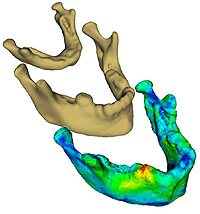 The Mathematical Modelling of Mandibular Metamorphosis (4M) project is aimed
at establishing models of the biological growth of human mandibles (jaw
bones) based on 3D CT (computed tomography) scans and clinically identified
landmarks. The project is a collaboration between both national and
international centres of expertise. The mathematical derived models aid in
the understanding of both mandibular growth and tooth eruption, and are
targeted for prediction, simulation, and analysis of biological and medical
processes. The ultimate goal of the research is, through improved understanding of
normal and abnormal craniofacial morphogenesis (including tooth eruption),
to improve treatment diagnosis, planning, and outcome in patients with
severe congenital craniofacial malformations. more
The Mathematical Modelling of Mandibular Metamorphosis (4M) project is aimed
at establishing models of the biological growth of human mandibles (jaw
bones) based on 3D CT (computed tomography) scans and clinically identified
landmarks. The project is a collaboration between both national and
international centres of expertise. The mathematical derived models aid in
the understanding of both mandibular growth and tooth eruption, and are
targeted for prediction, simulation, and analysis of biological and medical
processes. The ultimate goal of the research is, through improved understanding of
normal and abnormal craniofacial morphogenesis (including tooth eruption),
to improve treatment diagnosis, planning, and outcome in patients with
severe congenital craniofacial malformations. more
Contact:
Rasmus Larsen,
Klaus Baggesen Hilger
Automated Segmentation and Analysis of Cardiac MRI
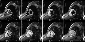
 Magnetic resonance imaging (MRI) has been shown to be an accurate and precise technique to assess cardiac volumes and function in a non-invasive manner and is generally considered to be the current gold standard for cardiac imaging. Measurement of ventricular volumes, muscle mass and function is based on determination of the left-ventricular endocardial and epicardial borders. Since manual border detection is laborious, automated segmentation is highly desirable as a fast, objective and reproducible alternative. This project develops methods for automated segmentation and will thus enhance comparability between and within cardiac studies and increase accuracy by allowing acquisition of thinner MRI-slices.
more
Magnetic resonance imaging (MRI) has been shown to be an accurate and precise technique to assess cardiac volumes and function in a non-invasive manner and is generally considered to be the current gold standard for cardiac imaging. Measurement of ventricular volumes, muscle mass and function is based on determination of the left-ventricular endocardial and epicardial borders. Since manual border detection is laborious, automated segmentation is highly desirable as a fast, objective and reproducible alternative. This project develops methods for automated segmentation and will thus enhance comparability between and within cardiac studies and increase accuracy by allowing acquisition of thinner MRI-slices.
more
Contact:
Mikkel B. Stegmann
Motion-compensation of Cardiac Perfusion MRI
 Within the last decade magnetic resonance imaging has been proven able to assess myocardial perfusion in an accurate and safe manner. While scanning times have improved drastically, the amount of manual post-processing remains to render the method prohibitive to clinical practice. A major part of this manual labour is spent by marking up points of correspondence on the myocardium, thus enabling compensation of any motion during a perfusion sequence. This project aims at replacing the tedious and error prone labour with an automatic image analysis method, which provides a structured way of collecting and applying expert knowledge given by medical doctors into a learning-based framework.
more
Within the last decade magnetic resonance imaging has been proven able to assess myocardial perfusion in an accurate and safe manner. While scanning times have improved drastically, the amount of manual post-processing remains to render the method prohibitive to clinical practice. A major part of this manual labour is spent by marking up points of correspondence on the myocardium, thus enabling compensation of any motion during a perfusion sequence. This project aims at replacing the tedious and error prone labour with an automatic image analysis method, which provides a structured way of collecting and applying expert knowledge given by medical doctors into a learning-based framework.
more
Contact:
Mikkel B. Stegmann
3D Shape Analysis of The Craniofacial Anomaly in Children With Cleft Lip and Palate
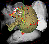 This project develops methods for extraction and analysis of the shape and size of the human skull in infancy and adolescence, and is carried out at the joint 3D-Laboratory (3D-Lab) of Copenhagen University Hospital, School of Dentistry, University of Copenhagen and Informatics and Mathematical Modelling, Technical University of Denmark. The methods are applied to three-projection x-ray images, plaster casts of palatal impressions and three-dimensional scans of children with cleft lip and palate. Reliable and detailed (semi-) automatic 3D point-to-point correspondence across a population of shapes is achieved using deformable models. Statistical methods are applied in order to analyze the shape and size variation within groups of children, as well as in order to discern between different types of treatment and study temporal evolution.
more
This project develops methods for extraction and analysis of the shape and size of the human skull in infancy and adolescence, and is carried out at the joint 3D-Laboratory (3D-Lab) of Copenhagen University Hospital, School of Dentistry, University of Copenhagen and Informatics and Mathematical Modelling, Technical University of Denmark. The methods are applied to three-projection x-ray images, plaster casts of palatal impressions and three-dimensional scans of children with cleft lip and palate. Reliable and detailed (semi-) automatic 3D point-to-point correspondence across a population of shapes is achieved using deformable models. Statistical methods are applied in order to analyze the shape and size variation within groups of children, as well as in order to discern between different types of treatment and study temporal evolution.
more
Contact:
Tron Darvann
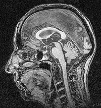
Corpus Callosum Morphometry
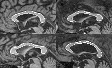 Corpus callosum is the nervous tissue that connects the two cerebral hemispheres of the human brain. Many neurological studies indicate that the size and shape of the corpus callosum are related to gender, age, neurodegenerative diseases et cetera. The gold standard for such morphometry studies is magnetic resonance imaging, which allows acquisition of accurate images of the anatomy (and function) of the brain. However, obtaining manual tracings of the corpus callosum is both time-consuming, error-prone and operator dependent. Instead, this project aims at replacing this task with automated and efficient methods eliminating subjectivity.
more
Corpus callosum is the nervous tissue that connects the two cerebral hemispheres of the human brain. Many neurological studies indicate that the size and shape of the corpus callosum are related to gender, age, neurodegenerative diseases et cetera. The gold standard for such morphometry studies is magnetic resonance imaging, which allows acquisition of accurate images of the anatomy (and function) of the brain. However, obtaining manual tracings of the corpus callosum is both time-consuming, error-prone and operator dependent. Instead, this project aims at replacing this task with automated and efficient methods eliminating subjectivity.
more
Contact:
Mikkel B. Stegmann
Statistical Shape Analysis of the Human Ear Canal
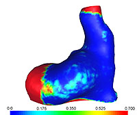 Hearing aids come in a number of different styles. The smallest of
these styles is called CIC (completely in the canal) and it has a
number of attractive properties. First of all the small size is
cosmetically appealing because a well-produced CIC is as good as
invisible in-situ. Secondly, the CIC has some acoustic advantages.
The shell of a CIC must have size and shape that allows it to contain a
microphone, an amplifier, a loudspeaker, and battery. On the other
hand it is desirable to minimise the size of the final hearing aid.
Today the design of components is based on intuition and skill and
not on a systematic description of the ear canal. It is acknowledged
that systematic knowledge of the geometry of ear canals and the
variation thereof potentially could be extremely helpful in the
mechanical design of new components for hearing aids. It is obvious
that the systematic description of the variation of the shape of the
ear canal must be done using statistical methods.
more
Hearing aids come in a number of different styles. The smallest of
these styles is called CIC (completely in the canal) and it has a
number of attractive properties. First of all the small size is
cosmetically appealing because a well-produced CIC is as good as
invisible in-situ. Secondly, the CIC has some acoustic advantages.
The shell of a CIC must have size and shape that allows it to contain a
microphone, an amplifier, a loudspeaker, and battery. On the other
hand it is desirable to minimise the size of the final hearing aid.
Today the design of components is based on intuition and skill and
not on a systematic description of the ear canal. It is acknowledged
that systematic knowledge of the geometry of ear canals and the
variation thereof potentially could be extremely helpful in the
mechanical design of new components for hearing aids. It is obvious
that the systematic description of the variation of the shape of the
ear canal must be done using statistical methods.
more
Contact:
Rasmus R. Paulsen
Efficient Analysis and Synthesis of 2D Face Images
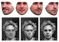 Generative models capable of synthesising photo-realistic images of objects have been shown to be powerful tools for image interpretation. Especially the Active Appearance Model framework have proven very capable within face segmentation, face analysis and face recognition. In this project we explore composite appearance representations and wavelet compression schemes et cetera, to obtain accurate and efficient models for analysis and synthesis of planar face images. Given this 2D problem, we aim at unleashing the power of contemporary graphics board to lessen the otherwise substantial computational load on the main CPU and resulting in models capable of tracking human faces in real-time. Applications include: biometric security, digital puppeteering, lip-reading, eye-tracking, binding et cetera. A subset of our face database has been put in the public domain for the benefit of future research.
more
Generative models capable of synthesising photo-realistic images of objects have been shown to be powerful tools for image interpretation. Especially the Active Appearance Model framework have proven very capable within face segmentation, face analysis and face recognition. In this project we explore composite appearance representations and wavelet compression schemes et cetera, to obtain accurate and efficient models for analysis and synthesis of planar face images. Given this 2D problem, we aim at unleashing the power of contemporary graphics board to lessen the otherwise substantial computational load on the main CPU and resulting in models capable of tracking human faces in real-time. Applications include: biometric security, digital puppeteering, lip-reading, eye-tracking, binding et cetera. A subset of our face database has been put in the public domain for the benefit of future research.
more
Contact:
Mikkel B. Stegmann
3D Facial Surface Appearance Models
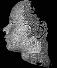 This project is concerned with modelling and analysis of 3D
surface appearance models of human faces and fitting these models
to 2D facial images.
In collaboration with the 3D-Laboratory, School of Dentistry,
University of Copenhagen a 3D face database is compiled using their
Minolta Vivid 900 shape-and-texture scanner.
The model building includes semi-automated registration of shape-and-texture
for all faces in the database, alignment and decomposition into an
appearance model. The model is used to segment 2D facial images.
This project is concerned with modelling and analysis of 3D
surface appearance models of human faces and fitting these models
to 2D facial images.
In collaboration with the 3D-Laboratory, School of Dentistry,
University of Copenhagen a 3D face database is compiled using their
Minolta Vivid 900 shape-and-texture scanner.
The model building includes semi-automated registration of shape-and-texture
for all faces in the database, alignment and decomposition into an
appearance model. The model is used to segment 2D facial images.
Contact:
Rasmus Larsen
Facial and Retinal Analysis, Modelling and Estimation
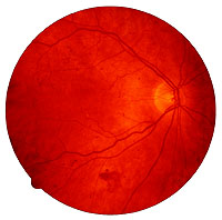 The FRAME project aims at 3D reconstruction of biological form, applied to retina and human faces. In the case of 3D reconstruction of the retina, the goal is to develop algorithms, which are able to distinguish between variations in the healthy eye versus the deformations present in the abnormal eye. In the part involving modelling of the human face, 3D scans of form and texture will be used to estimate a 3D Active Appearance Model (AAM). Modelling of the shape, texture and dynamic will be done on the basis of a 3D face database, which is under construction in collaboration with the 3D laboratory at the dental institute of Copenhagen. The project will include methods from modern multivariante statistics to construct realistic models.
Contact:
Brian Lading
Active Appearance Models for Segmentation of Bone Structures
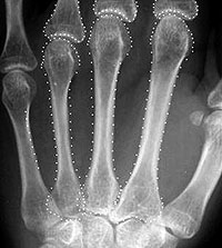 This project seeks to obtain better accuracy, precision and robustness of the segmentation of bones in projection radiographs and MR images. This is pursued by building prior models of the shape and intensity distributions for the bones in the framework of Active Appearance Models (AAM). There is plenty of room for improvement, and current issues are:
1) How to obtain accurate reconstruction of the bone edges
2) Understanding the occasional errors that AAM commits
3) Automating the constructing of training sets for AAM
4) Adapting AAM to partially corrupted images
5) Use of AAM in serial images for clinical trials: How to obtain better precision and better sensitivity to changes
more
Contact:
Hans Henrik Thodberg
Minimum Description Length Shape and Appearance Models
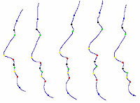 This project develops methods and software to place landmarks on a set of contours in a consistent and automated fashion. As guiding principle we use Ockham's Razor in the guise of the Minimum Description Length (MDL) formalism. It is believed that more correct placement of the marks leads to a more compact statistical shape or appearance model, and thus the mark positions are gradually improved using compactness as objective function.
The task is formulated as unsupervised learning using a single labelled example, and a generalisation to image segmentation is investigated.
A Matlab open source toolbox has been released, which successfully places landmarks on 2D curves.
more
This project develops methods and software to place landmarks on a set of contours in a consistent and automated fashion. As guiding principle we use Ockham's Razor in the guise of the Minimum Description Length (MDL) formalism. It is believed that more correct placement of the marks leads to a more compact statistical shape or appearance model, and thus the mark positions are gradually improved using compactness as objective function.
The task is formulated as unsupervised learning using a single labelled example, and a generalisation to image segmentation is investigated.
A Matlab open source toolbox has been released, which successfully places landmarks on 2D curves.
more
Contact:
Hans Henrik Thodberg
Automated Characterization and Recognition of 2D and 3D Brain Structure in MRI for Diagnostic Support
 This project is concerned with accurate characterization and
segmentation of brain structures in magnetic resonance imagery and
relation of derived parameters to clinical and cognitive
performance data. This is relevant for individual diagnosis of
brain diseases such as dementia and epilepsy, surgical planning,
and for population studies of the effect of particular drugs. The
project is a collaboration between
Informatics and Mathematical
Modelling, DTU and the
MR Department at Hvidovre University
Hospital.
Overall this project is part of a trend for image analysis at IMM to address vision system optimisation. The optimisation is made over imaging device, image processing, image analysis, and decision support system.
This project is concerned with accurate characterization and
segmentation of brain structures in magnetic resonance imagery and
relation of derived parameters to clinical and cognitive
performance data. This is relevant for individual diagnosis of
brain diseases such as dementia and epilepsy, surgical planning,
and for population studies of the effect of particular drugs. The
project is a collaboration between
Informatics and Mathematical
Modelling, DTU and the
MR Department at Hvidovre University
Hospital.
Overall this project is part of a trend for image analysis at IMM to address vision system optimisation. The optimisation is made over imaging device, image processing, image analysis, and decision support system.
Contact:
Rasmus Larsen
Novelty Detection In Dermatological Images
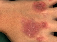 This project is focused upon two important topics in pattern recognition of image data, namely spectral texture models and dynamic image analysis. Current texture models generally only deal with one of the four fundamental issues: scale, colour, morphology, and non-stationarity. The work is aimed at constructing texture models incorporating all four issues, developing simulation and estimation algorithms for these models, and applying them for image interpretation in the application domain. The collection of images over time is analysed in order to find changes in a multivariate fashion.
Contact:
Bjarne K. Ersbĝll
Apparatus for Skin Imaging and Skin Image Representation
 Nowadays, the medical tracking of dermatological diseases is imprecise. The main reason is the lack of suitable objective methods to evaluate the lesion. Doctors visually asses the lesion and make scorings and journal notes of the condition. These notes and perhaps some photographs are presently the only memory of what the lesion looked like at the corresponding patient visit. The purpose of this project is to develop an image system that allows tracking the lesion in an objective way. Project tasks include:
1) to get accurate dermatological images using a high tech image system and
to extract the lesion using image analysis tools.
2) development of mathematical and statistical models to summarize the
lesion and track the evolution.
3) analysis of multispectral images.
Nowadays, the medical tracking of dermatological diseases is imprecise. The main reason is the lack of suitable objective methods to evaluate the lesion. Doctors visually asses the lesion and make scorings and journal notes of the condition. These notes and perhaps some photographs are presently the only memory of what the lesion looked like at the corresponding patient visit. The purpose of this project is to develop an image system that allows tracking the lesion in an objective way. Project tasks include:
1) to get accurate dermatological images using a high tech image system and
to extract the lesion using image analysis tools.
2) development of mathematical and statistical models to summarize the
lesion and track the evolution.
3) analysis of multispectral images.
Contact:
David Delgado Gomez